Part 2 in this series describes the ligaments and capsules in the shoulder. We focus on the glenohumeral joint (GHJ), but will touch upon some of the other functionally important structures.
The Shoulder Ligaments
Ligaments are band like structures that typically attach one end of the joint to the other. Its purpose is to stabilise the joint throughout its whole range of motion. It ensures the joint never dislocates or pops in and out. They are passive structures. This means that they do not require any activation in order for them to perform its function. Unlike muscles they usually do not have a blood supply and rely on its mechanical properties to do its job[1].
Figure 1 below shows the GH ligaments which together form part of the GH capsule containing the GH joint. It also shows 3 other ligaments which are key in the functionality of the shoulder.
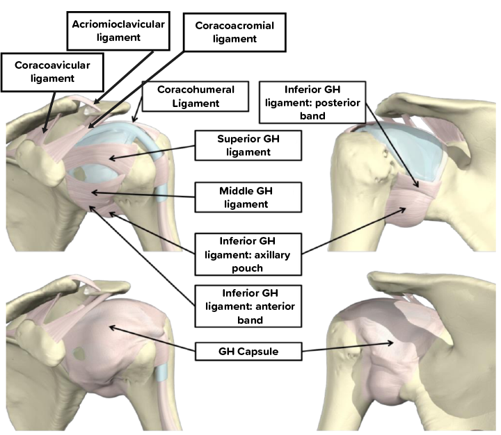
Figure 1
The GH capsule is made up of capsular structure that completely encloses the GH joint. At one end it is attached around the edge of the humeral head sometimes referred to as the anatomical neck and at the other end attached to the complete surrounding of the glenoid. It is the GH ligaments that give the capsule its strength, as alone it cannot hold the joint together.
Function of Shoulder Ligaments
The most basic function of ligaments is to prevent the joint from pulling apart, dislocating or from subluxation. In addition there is a theory, which is now widely accepted as fact in biomechanics and biology; there are mechanoreceptors within the ligaments [1, 2]. Mechanoreceptors can be thought of as stretch sensors. The sensor picks up the changes in stretch of whatever it is embedded into and sends a signal as appropriate. In the case of mechanoreceptors in the GH ligaments, they sense how much the ligaments have stretched and send a signal to the nervous system. If the ligaments are stretching too much the nervous system would react by activating the rotator cuff muscles to ensure the joint remains stable.
What Could Go Wrong?
When the ligaments themselves become fibrous, or tear, or become damaged in anyway, its mechanical properties change. The function of the ligaments relies heavily on its mechanical properties, more so its resistance and behaviour to tension. If the ligaments become more stiff they could tear much more easily. If they become too lax they are not taught enough to provide the resistance needed to prevent any form of instability. In both instances, the behaviour of the mechanoreceptors change as well. This means that the feedback loop with the rotator cuff muscles are also affected. Leading to a further loss of stability in the shoulder.
Losing stability isn’t necessarily the only result of changes in the shoulder. If the ligaments do become more stiff but not too stiff to tear easily, can cause the shoulder to loose its main purpose; a wide range of motion. It would not just be a physical loss in range of motion, but it would very likely be accompanied by significant pain. Ever twisted your ankle? Do you remember the pain/ That pain is caused by over-stretching of the ligaments. Now, imagine the ligaments are now much more stiff. The point at which they become over stretched is now much closer to its normal range of function. So you would feel the pain earlier, and very likely significantly more so than if the ligament was normal.
But Ligaments Are Not Enough to Stabilise the Shoulder
If ligaments are not enough to stabilise the shoulder, then what is? What is the primary source of stability? Part 3 and the final part of this series discusses the muscles that span the shoulder joint. The rotator cuff muscles will be discussed in depth as this is one of the main sources of problems that patients experience with.